
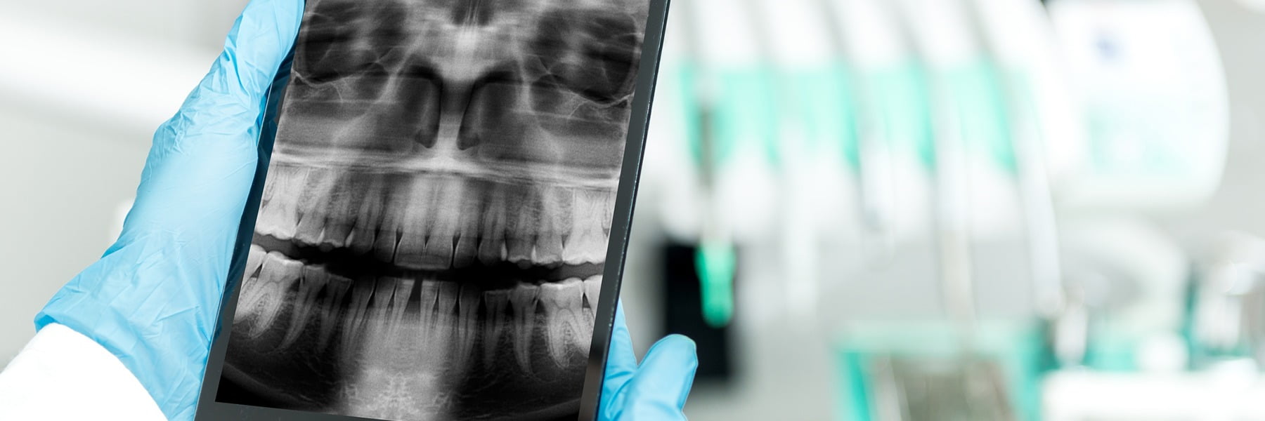
Dental X-rays allow your dentist to see areas of your teeth, roots and jawbone that cannot be examined simply by looking inside your mouth. They can reveal early tooth decay between teeth, infections around tooth roots, bone loss caused by gum disease, impacted wisdom teeth and many other problems that may otherwise remain hidden.
However, this does not mean that everybody needs X-rays at every dental appointment. In the UK, dental X-rays should only be taken when there is a clinical reason for the image and the expected diagnostic benefit justifies the very small radiation exposure.
At Glow Dental in Battersea, we use modern digital dental radiography and 3D imaging to support diagnosis and treatment planning. Where appropriate, we can also show you your images and explain what we can see before discussing any recommended treatment.
What Can a Dental X-Ray Show?
Different types of dental X-ray reveal different structures, but they can help dentists identify problems that are difficult or impossible to see during a normal visual examination.
Dental X-rays may help identify:
- Tooth decay between teeth
- Decay underneath or around existing restorations
- Infection around the root of a tooth
- Bone loss associated with periodontal disease
- Impacted or unerupted teeth
- Wisdom-tooth position
- Root shape and anatomy
- Problems associated with previous root canal treatment
- Some cysts and other changes affecting the jaws
- Developing permanent teeth in children
- Missing or additional teeth
- Bone available for dental implants
- The position of teeth before orthodontic treatment
An X-ray is therefore not simply a picture of a cavity. It provides information about structures that lie beneath the visible surface.
Can Dental X-Rays Detect Cavities?
Yes. Dental X-rays are particularly useful for detecting tooth decay in areas that cannot easily be inspected directly.
One of the most common examples is decay between neighbouring teeth.
When two teeth touch closely together, your dentist cannot necessarily see the entire contact area during an examination. A bitewing X-ray can show whether minerals have been lost from the enamel or dentine between the teeth.
X-rays can also sometimes reveal decay:
- Around the edges of existing fillings
- Beneath crowns
- On root surfaces
- In areas that are difficult to examine directly
Not every tiny cavity is automatically visible on an X-ray, particularly in its earliest stages. Dentists therefore combine radiographs with a clinical examination rather than relying on an image alone.
If tooth decay has progressed enough to create a cavity, treatment such as a tooth-coloured filling may be necessary.
Can Dental X-Rays Show an Infection or Abscess?
They often can.
If the nerve inside a tooth has become infected or died, inflammation can eventually affect the bone around the end of the root.
On a dental X-ray, this can sometimes appear as a darker area around the root tip.
Radiographs can therefore help dentists assess:
- Dental abscesses
- Infection around a root
- The condition of an infected tooth
- Previous root canal treatment
- The anatomy of the roots before treatment
However, an infection does not always create an immediately visible X-ray change. Very early inflammation may be present before significant changes develop within the surrounding bone.
Your dentist therefore considers your symptoms, examination findings and X-rays together.
If an infected tooth can be saved, root canal treatment may be used to remove infection from inside the tooth.
Can Dental X-Rays Show Gum Disease?
Dental X-rays can show one of the most important consequences of advanced gum disease: loss of the bone supporting the teeth.
Healthy teeth are surrounded by supporting jawbone. When periodontitis progresses, inflammation can gradually destroy some of this bone.
Radiographs can help a dentist or periodontist assess:
- The height of bone around the teeth
- The pattern of bone loss
- Whether certain areas are more severely affected
- Changes over time when previous images are available
However, an X-ray cannot replace an examination of the gums. Your dentist also needs to assess bleeding, periodontal pocket depths, gum recession, tooth mobility and plaque levels.
If more advanced periodontal disease is present, Glow Dental provides specialist periodontal treatment.
Can Dental X-Rays Show Wisdom Teeth?
Yes. A panoramic dental X-ray — commonly called an OPG — can provide an overview of all the teeth and both jaws.
This can be useful for assessing wisdom teeth that are:
- Partially erupted
- Completely buried within the jaw
- Growing at an angle
- Close to neighbouring teeth
For lower wisdom teeth, the dentist may also need to consider how closely the roots lie to important nerves within the lower jaw.
If a two-dimensional X-ray does not provide enough information in a complex case, a 3D CBCT scan may occasionally be recommended.
Can Dental X-Rays Show a Cracked Tooth?
Sometimes, but not always.
This is an important limitation of dental radiography.
Many tooth cracks are extremely fine or run in a direction that is difficult to see on a conventional X-ray.
A dentist may therefore diagnose a cracked tooth using a combination of:
- Your symptoms
- A clinical examination
- Bite testing
- Magnification
- Transillumination
- Dental X-rays
An apparently normal X-ray does not necessarily prove that a painful tooth is not cracked.
Can Dental X-Rays Show Oral Cancer?
Dental X-rays are not the main screening test for mouth cancer and cannot independently diagnose oral cancer.
Mouth cancer screening primarily involves examining the lips, tongue, gums, cheeks, floor and roof of the mouth and other visible soft tissues.
Radiographs may sometimes show changes affecting the jawbone or other structures that require further investigation, but a suspicious mouth lesion generally requires clinical assessment and, where indicated, specialist referral and biopsy.
At Glow Dental, mouth cancer screening forms part of routine dental examinations.
Can Dental X-Rays Show Problems Under Crowns and Fillings?
They can reveal some problems that would otherwise remain hidden.
For example, an X-ray may help identify:
- Decay around the margin of a restoration
- Decay beneath certain areas of a crown or filling
- Infection affecting the root of a crowned tooth
- The condition of previous root canal treatment
- Changes in the bone supporting the tooth
However, some dental materials appear very dense on X-rays and can obscure the tooth beneath them. Your dentist therefore combines radiographic information with a physical examination.
Can Dental X-Rays Show Developing Children’s Teeth?
Yes. X-rays can be particularly useful when a dentist needs to assess how a child’s permanent teeth are developing underneath the baby teeth.
They can show:
- Permanent teeth that have not yet erupted
- Missing permanent teeth
- Extra teeth
- Teeth developing in unusual positions
- Dental decay between baby or permanent teeth
- Root development
However, children do not automatically need X-rays simply because they are young.
As with adults, radiographs should only be taken when the dentist believes the information will help with diagnosis or treatment.
You can learn more about looking after developing teeth through our children’s dentistry service.
What Are the Different Types of Dental X-Ray?
The term “dental X-ray” actually covers several different types of image.
Bitewing X-Rays
Bitewings normally show the crowns of the upper and lower back teeth together.
They are particularly useful for:
- Detecting decay between teeth
- Monitoring existing fillings
- Assessing the level of supporting bone
You gently bite onto a small holder while the image is taken.
Periapical X-Rays
A periapical X-ray shows the whole tooth, from the crown down to the tip of the root and surrounding bone.
It can be useful when investigating:
- Toothache
- Dental abscesses
- Root problems
- Dental trauma
- Root canal treatment
Panoramic X-Ray or OPG
An OPG provides a broad two-dimensional image of both jaws and most or all of the teeth.
The machine rotates around your head while you stand or sit still.
An OPG may be useful for:
- Wisdom-tooth assessment
- Orthodontic planning
- Looking at several missing or unerupted teeth
- Assessing the jaws more generally
- Some implant and surgical assessments
Cephalometric X-Ray
A cephalometric radiograph provides a side-on image of the head and jaws.
It is mainly used as part of orthodontic assessment to examine the relationship between the teeth, jaws and facial structures.
Glow Dental provides both Invisalign and specialist orthodontic care, with imaging selected according to the individual treatment plan.
CBCT or 3D Dental Scan
CBCT stands for cone beam computed tomography.
Unlike an ordinary dental X-ray, which produces a flat two-dimensional picture, CBCT generates a three-dimensional image of the teeth and jaws.
This can provide detailed information about:
- Jawbone dimensions
- Tooth roots
- Nerves
- Sinuses
- Impacted teeth
- Complex root canal anatomy
- Potential dental implant sites
At Glow Dental, 3D imaging and CT technology can be used to support complex treatment planning, including dental implant treatment.
Is a CBCT Scan the Same as a Normal Dental X-Ray?
No.
CBCT provides considerably more three-dimensional information, but it also generally involves a higher radiation dose than a small conventional dental X-ray.
It should therefore not be taken simply because more detail might be interesting.
It is used when the additional 3D information is expected to make a meaningful difference to diagnosis or treatment planning.
For example, Guy’s and St Thomas’ NHS Foundation Trust explains that dental CBCT can show the exact structure and shape of the jawbone and may be used when assessing patients for dental implants.
Are Dental X-Rays Safe?
Dental X-rays use ionising radiation, so they are not completely risk-free. However, the radiation doses involved in ordinary dental radiography are very low.
UK radiation-protection regulations require every exposure to be clinically justified and the dose to be kept as low as reasonably practicable while still producing the information needed.
For context, UK Government patient-dose information gives typical effective doses of approximately:
- A single bitewing dental X-ray: less than 0.01 mSv
- A panoramic dental X-ray: around 0.01 mSv
These are each broadly comparable with only a small amount of the natural background radiation everybody receives simply by living in the UK.
The exact dose varies according to the machine, patient, imaging technique and type of examination.
Are Digital Dental X-Rays Safer Than Traditional Film?
Modern digital systems can allow high-quality dental images to be produced efficiently and with low radiation doses.
However, statements such as “digital X-rays always use 90% less radiation” are too simplistic because the actual dose reduction depends on the type of film or digital sensor, equipment and exposure settings being compared.
The more useful reassurance is that modern dental radiography is performed under strict UK radiation-protection rules designed to minimise unnecessary exposure.
Glow Dental uses digital radiography as part of its diagnostic technology.
Do You Need a Lead Apron for Dental X-Rays?
Not routinely.
Older images of dental radiography often show every patient wearing a large lead apron. Current UK radiation-protection guidance states that there is no justification for routine lead-apron use during ordinary dental radiography.
This is because modern dental X-ray beams are carefully restricted to the area being examined and the radiation dose is already very low.
A thyroid collar is also not automatically required for every image. It may be used in selected circumstances if the thyroid could lie within the primary X-ray beam and a radiation-protection expert advises that it is appropriate.
Are Dental X-Rays Safe During Pregnancy?
Yes, dental X-rays can be taken during pregnancy when they are clinically necessary.
The X-ray beam used for dental radiography is directed towards the mouth and jaws rather than the abdomen.
Guy’s and St Thomas’ NHS Foundation Trust states that dental X-rays are considered safe during pregnancy and can be taken when required for diagnosis or treatment.
You should still tell your dentist if you are pregnant or think you may be pregnant so that this can be considered as part of your care.
Non-urgent imaging that would not affect your immediate treatment may sometimes be postponed simply because there is no clinical reason to take it at that time — not because ordinary dental radiographs are expected to harm the pregnancy.
How Much Radiation Is There in a Dental X-Ray?
The answer depends on the image.
Small intraoral dental X-rays expose the patient to very low doses.
UK Government guidance estimates a single bitewing at less than 0.01 millisieverts (mSv) and a panoramic image at approximately 0.01 mSv.
For comparison, natural radiation is continuously present in our environment from the ground, atmosphere, food and cosmic radiation.
A CBCT scan has a higher dose than a simple bitewing or panoramic X-ray because it gathers considerably more information.
Guy’s and St Thomas’ estimates that a dental CBCT of the jaw can be comparable to approximately 6–30 days of normal background radiation, depending on the scan.
This is why CBCT is reserved for situations where the additional three-dimensional information is clinically useful.
How Often Should You Have Dental X-Rays?
There is no rule that everybody needs dental X-rays every six months, every year or at every check-up.
The decision should be based on your individual clinical need.
A dentist may consider factors such as:
- Your current examination findings
- Your history of tooth decay
- Whether you have new symptoms
- Your gum health
- Your age
- Previous X-rays
- Existing restorations
- Whether teeth are developing or erupting
- Any treatment being planned
A patient who repeatedly develops decay may require radiographs more frequently than an adult who has remained decay-free for years.
Similarly, a patient arriving with severe toothache may require an X-ray even if their last routine images were taken relatively recently.
Do You Need Dental X-Rays at Every Check-Up?
No.
Your dental examination and your X-ray schedule are related but are not the same thing.
Your dentist first assesses your mouth and reviews your dental history.
An X-ray is recommended only if it is expected to provide information that could influence:
- Diagnosis
- Monitoring
- Treatment planning
If an existing recent X-ray already provides the information required, there may be no reason to repeat it.
Can You Have Too Many Dental X-Rays?
Ionising-radiation exposure is cumulative, which is why dentists should avoid unnecessary radiographs.
This does not mean you should refuse a clinically justified dental X-ray because you have had previous ones.
Instead, UK practice follows two key principles:
Justification: there should be a valid clinical reason for the X-ray.
Optimisation: the exposure should be kept as low as reasonably practicable while producing an image good enough to answer the clinical question.
If you are unsure why an X-ray has been recommended, ask your dentist what they are looking for and how the result may influence your treatment.
Is a Dental X-Ray Painful?
No. The X-ray exposure itself does not hurt.
For a small intraoral X-ray, a sensor or image receptor is positioned inside the mouth while the X-ray is taken.
Some people find the holder slightly uncomfortable, particularly if they:
- Have a small mouth
- Have sensitive tissues
- Have a strong gag reflex
- Have painful or swollen teeth
The image itself is normally captured in a fraction of a second.
Panoramic X-rays and CBCT scans do not normally require a sensor to be positioned deep inside the mouth, which some patients find more comfortable.
What if You Have a Strong Gag Reflex?
Tell the dental team before an intraoral X-ray is attempted.
Small changes in sensor positioning, breathing techniques and taking the image efficiently can make the process easier.
In some situations, another type of image may be clinically appropriate, although an alternative should only be chosen if it can provide the information the dentist actually needs.
How Long Does a Dental X-Ray Take?
Dental radiographs are generally very quick.
A small bitewing or periapical X-ray may take only a few minutes to position and complete, with the actual exposure lasting only a tiny fraction of that time.
A panoramic OPG usually takes only a few minutes, while the actual scan around the head is brief.
CBCT scanning is also relatively quick, although additional time may be needed to position the patient correctly and analyse the three-dimensional images.
With digital radiography, images are available almost immediately for the dentist to review.
Do You Get the Results Straight Away?
For most routine digital dental X-rays, yes.
The dentist can normally view the image on a computer screen immediately.
This allows them to:
- Check the image quality
- Explain relevant findings
- Compare with previous X-rays where available
- Discuss treatment options
At Glow Dental, photographs, scans and X-rays can be used to help explain findings so that patients can understand why treatment is being recommended.
Dental X-Ray vs Digital Scan: What’s the Difference?
These are often confused.
A dental X-ray uses ionising radiation and allows the dentist to see structures such as tooth roots and bone.
An intraoral digital scan uses an optical scanner to create a three-dimensional model of the visible surfaces of your teeth and gums.
An optical scan does not replace an X-ray because it cannot see through enamel or bone.
At Glow Dental, both technologies may be used because they answer different clinical questions.
Why Are X-Rays Important Before Dental Implants?
Dental implant treatment requires detailed knowledge of the jawbone and surrounding anatomy.
Before placing an implant, the dentist may need to assess:
- The height and width of available bone
- The position of nearby roots
- Nerves in the lower jaw
- The maxillary sinus in the upper jaw
- The proposed implant angle and position
A conventional X-ray may provide useful initial information, but a CBCT scan is often valuable where precise three-dimensional assessment is required.
Glow Dental uses digital scanning and 3D imaging as part of planning suitable dental implant cases.
Why Are X-Rays Important Before Root Canal Treatment?
Root canal treatment involves structures inside the tooth that cannot be seen directly from the outside.
X-rays help the dentist or endodontist assess:
- The number and shape of the roots
- The approximate root canal anatomy
- Infection around the root tips
- Previous root canal fillings
- Changes in surrounding bone
Images may also be taken during and after treatment to help assess the procedure.
More complex cases can be managed through Glow Dental’s specialist root canal service.
How Much Does a Dental X-Ray Cost?
Private dental X-ray costs vary between practices and depend on the type of image required.
At Glow Dental, our current published fees include:
- New patient examination, including small X-rays where clinically required: £90
- Panoramic OPG X-ray: £95
More specialised imaging, such as CBCT, depends on the type and purpose of the scan. Your dentist will explain any additional imaging required and the associated cost before treatment.
Fees can change, so check our current dental fee guide for the latest prices.
Can You Refuse a Dental X-Ray?
You are entitled to ask why an X-ray is being recommended and to make an informed decision about your care.
Your dentist should be able to explain:
- What type of X-ray is proposed
- Why it is needed
- What information they expect it to provide
- How that information may affect your treatment
You can decline an X-ray. However, if an image is necessary to diagnose a problem or carry out treatment safely, your dentist may not be able to provide that particular treatment without the required diagnostic information.
What Can Dental X-Rays Not Show?
Dental X-rays are extremely useful, but they do not show everything.
For example, they may not reliably show:
- Very early tooth decay
- Some fine cracks
- The true depth of every cavity
- Every soft-tissue condition
- Mouth cancer by themselves
- Whether a tooth is actually painful
This is why diagnosis is based on a combination of your symptoms, medical and dental history, clinical examination and appropriate imaging.
Frequently Asked Questions About Dental X-Rays
Can a dental X-ray show every cavity?
No. X-rays are very useful for detecting decay, particularly between teeth, but very early lesions and some cavities may not be clearly visible. Clinical examination remains essential.
Can an X-ray show a tooth infection?
Often, especially once the infection has caused changes in the bone around the root. Very early infections may not yet produce obvious X-ray changes.
Can dental X-rays detect gum disease?
They can demonstrate supporting-bone loss associated with periodontitis, but the gums also need to be examined and periodontal measurements taken.
Can an X-ray show a cracked tooth?
Sometimes, but many cracks are too fine or positioned in a way that makes them difficult to see on a conventional dental X-ray.
Are dental X-rays safe?
They involve ionising radiation, so the exposure is not literally zero risk, but conventional dental X-ray doses are extremely low. UK regulations require exposures to be justified and optimised.
How much radiation is in a bitewing X-ray?
UK Government patient-dose guidance lists a typical single bitewing exposure as less than 0.01 mSv, comparable with less than about a day and a half of average natural background radiation.
Are dental X-rays safe during pregnancy?
Yes, when clinically necessary. The dental X-ray beam is directed at the mouth and jaw rather than the developing baby.
Do children need dental X-rays?
Only when clinically justified. They can be useful for detecting decay and monitoring tooth development, but children should not have X-rays simply according to a fixed routine.
Do I need an X-ray every six months?
No. X-ray frequency should be based on your individual risk, symptoms, previous images and treatment needs.
Do I need a lead apron?
Not routinely. Current UK guidance does not recommend automatic lead-apron use for ordinary dental radiography.
Is a CBCT scan dangerous?
CBCT involves more radiation than a simple dental X-ray but considerably less than many conventional medical CT scans. It should only be used when the extra three-dimensional information is clinically justified.
Do dental X-rays hurt?
No. The radiation exposure itself cannot be felt. A small sensor inside the mouth may cause temporary discomfort in some patients.
How quickly can the dentist see my X-ray?
Digital images are generally available immediately, allowing your dentist to review them during the appointment.
Dental X-Rays at Glow Dental in Battersea
At Glow Dental, dental X-rays are used when they can provide meaningful information about your oral health or help us plan treatment safely.
Our practice uses modern technology including:
- Digital dental radiography
- Panoramic imaging
- 3D scanning
- CT imaging
These technologies can support everything from routine diagnosis to more complex treatment such as root canal therapy, orthodontics and dental implant planning.
Most importantly, X-rays are not taken simply because you have arrived for a routine appointment. Your dentist considers your symptoms, previous dental history, clinical findings and existing images before deciding whether new radiographs are necessary.
A routine dental examination at Glow Dental allows us to assess your teeth, gums and oral tissues and determine whether any additional diagnostic imaging would be useful.
If you have toothache, swelling or another urgent problem, our emergency dental service can assess the problem and arrange appropriate imaging where required.
Contact Glow Dental in Battersea to arrange an appointment.
For independent information about safety and radiation exposure, you can also read the Guy’s and St Thomas’ NHS guide to dental X-rays.
Written by:
Glow Dental
Published:
Last updated: