
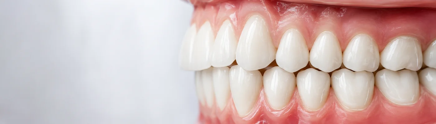
Premolars are permanent teeth positioned between the pointed canine teeth and the larger molar teeth at the back of the mouth.
Adults normally have eight premolars: four in the upper jaw and four in the lower jaw. Their raised chewing surfaces help crush and break down food before it reaches the molars for further grinding.
Premolars are also commonly called bicuspids because many have two main cusps. However, their anatomy can vary, and some premolars have more than two cusps.
How Many Premolars Do Adults Have? The Short Answer
An adult usually has eight permanent premolars.
These include:
- Four premolars in the upper jaw
- Four premolars in the lower jaw
There are two premolars in each quarter of the mouth:
- First premolar
- Second premolar
This means that the upper right, upper left, lower right and lower left parts of the mouth normally contain two premolars each.
Some adults may have fewer than eight because a premolar never developed, remained impacted, was removed for orthodontic reasons or was lost because of decay, gum disease, trauma or another dental problem.
Where Are Premolars Located?
Premolars sit immediately behind the canine teeth and in front of the first molars.
The order of the adult teeth from the centre of the mouth towards the back is generally:
- Central incisor
- Lateral incisor
- Canine
- First premolar
- Second premolar
- First molar
- Second molar
- Third molar or wisdom tooth
The position of the premolars allows them to provide a transition between the tearing action of the canines and the heavier grinding action of the molars.
Do Children Have Premolars?
Children do not have premolars in their primary or baby dentition.
Baby teeth include incisors, canines and primary molars. As the child develops, the primary molars become loose and are replaced by permanent premolars.
This differs from the first permanent molars, which emerge behind the baby teeth rather than replacing an existing tooth.
Permanent premolars commonly erupt between approximately 10 and 13 years of age, although timing varies between individual children and teeth. Glow Dental provides monitoring of tooth development and eruption as part of our children’s dentistry service.
When Do First and Second Premolars Erupt?
The permanent first and second premolars usually emerge during the later mixed-dentition stage, when a child has a combination of baby and adult teeth.
First premolars
The first premolars generally emerge before or around the same time as the second premolars. Their exact eruption age can vary between the upper and lower jaws.
Second premolars
The second premolars develop behind the first premolars and replace the second primary molars. They may emerge slightly later, although the eruption sequence varies.
A delayed premolar does not automatically indicate a problem. However, a dentist may recommend an examination or X-ray where:
- A baby molar remains in place longer than expected
- A premolar appears to be missing
- The tooth is erupting in an unusual position
- There is insufficient space in the dental arch
- Neighbouring teeth are moving into the available space
Regular dental examinations allow the development and position of the adult teeth to be monitored.
What Do Premolars Look Like?
Premolars are smaller than molars but have broader chewing surfaces than the canine teeth.
They generally have:
- A crown with raised cusps
- Grooves across the chewing surface
- A shape suited to crushing and breaking food
- One or more roots, depending on the individual tooth
Many premolars have two main cusps: one towards the cheek and one towards the tongue or palate. However, anatomy varies. Some lower second premolars, for example, can have three cusps.
The first premolar often appears slightly sharper, while the second premolar may have a broader, more rounded chewing surface.
Why Are Premolars Called Bicuspids?
The word “bicuspid” means a tooth with two cusps. This term is commonly used because many premolars have two main raised points on their chewing surfaces.
However, not every premolar has exactly two cusps. Because variations are possible, “premolar” is the more accurate general term for this group of teeth.
What Is the Function of Premolars?
Premolars have several important functions.
Crushing food
Their cusps press food against the opposing teeth, breaking it into smaller pieces before it reaches the molars.
Assisting with tearing
The first premolars can support the canine teeth when tearing firmer foods.
Supporting the molars
Premolars help transfer food towards the back teeth, where the broader molar surfaces carry out heavier grinding.
Distributing chewing forces
Premolars help distribute biting and chewing pressure across the dental arch rather than concentrating it on a smaller number of teeth.
Maintaining the bite
They also help maintain the position of neighbouring teeth and contribute to the height and stability of the bite.
What Is the Difference Between Premolars and Molars?
Premolars and molars are both back teeth used during chewing, but they differ in size, position and function.
| Feature | Premolars | Molars |
| Number in adults | Eight | Up to 12, including wisdom teeth |
| Position | Between canines and molars | At the back of the mouth |
| Main function | Crushing and breaking food | Heavier grinding and chewing |
| Primary dentition | Not present | Primary molars are present |
| Chewing surface | Smaller, usually with two or more cusps | Broader, with several cusps and grooves |
What Problems Can Affect Premolars?
Premolars are exposed to chewing pressure and can develop many of the same problems as other permanent teeth.
Tooth decay
Plaque and food can collect in the grooves on the chewing surfaces or between neighbouring teeth. Early decay may cause no symptoms, while deeper decay can lead to sensitivity, toothache or infection.
A small or moderate cavity may be restored with a dental filling.
Cracked cusps
A cusp may crack because of a large existing filling, heavy biting forces, tooth grinding or biting unexpectedly hard food.
Possible symptoms include:
- Sharp pain when biting
- Pain when releasing the bite
- Sensitivity to hot or cold
- Intermittent discomfort that is difficult to locate
Large or failing fillings
Older fillings can wear, fracture or develop leakage around their edges. The appropriate treatment depends on the size of the defect and the amount of healthy tooth remaining.
Where a direct filling may not provide enough protection, the dentist may consider an inlay or onlay or a dental crown.
Infection inside the tooth
If decay or a crack reaches the dental pulp, the tooth may become inflamed or infected. Symptoms can include persistent toothache, prolonged sensitivity, pain when biting or swelling.
Where the premolar remains restorable, root canal treatment may be used to remove damaged or infected tissue from inside the tooth.
Gum disease
Periodontal disease can damage the gums and bone supporting a premolar. Advanced disease may cause recession, deeper periodontal pockets and tooth mobility.
Tooth wear
Grinding, clenching or an uneven bite may wear down the cusps or place additional pressure on the tooth. Treatment depends on the amount of wear, the cause and whether symptoms are present.
Impacted or unerupted premolars
Occasionally, a premolar may fail to emerge normally because there is insufficient space, the tooth is displaced or another structure is blocking its path.
An examination and dental X-ray can help determine the position of the tooth and whether monitoring, orthodontic treatment or another procedure may be appropriate.
Can a Premolar Have Root Canal Treatment?
Yes. Root canal treatment can be performed on a premolar where the dental pulp is irreversibly inflamed, infected or dead and the tooth can still be restored.
Premolar root and canal anatomy varies. Some premolars have one root and one canal, while others have multiple roots or canal spaces. This can influence the complexity of treatment.
After root canal treatment, the tooth must be properly sealed and restored. Depending on the amount of healthy structure remaining, the dentist may recommend a filling, onlay or crown to protect it.
Why Are Premolars Sometimes Removed?
Dentists generally aim to preserve a healthy, restorable premolar. However, removal may be recommended in certain circumstances.
Orthodontic treatment
Premolars may sometimes be removed as part of a carefully planned orthodontic treatment where additional space is needed to manage significant crowding or correct the bite.
Extraction is not required for every orthodontic patient. The decision should follow a full assessment of:
- The amount and location of crowding
- The relationship between the upper and lower teeth
- The shape and size of the dental arches
- The position of the front teeth
- The patient’s facial profile
- Growth and development
- Alternative treatment options
Where removal is appropriate, the orthodontic appliances are then used to move other teeth into planned positions and manage the extraction spaces.
Extensive tooth decay
Extraction may be considered where decay has destroyed too much tooth structure for a predictable filling, onlay or crown.
Severe fracture
A premolar with a deep crack extending into the root may not be restorable.
Advanced gum disease
Severe loss of supporting bone can make a premolar very loose and reduce its long-term prognosis.
Persistent infection
Extraction may be recommended where infection cannot be predictably treated or where previous treatment has not resolved the problem and further treatment is unsuitable.
Does Premolar Extraction Hurt?
Premolar extraction is normally carried out using local anaesthetic. The area should therefore be numb during the procedure, although the patient may feel pressure or movement.
Some tenderness, swelling or minor bleeding can occur afterwards. The dentist will provide instructions covering cleaning, eating, pain relief and when to seek further advice.
Contact the practice if pain becomes increasingly severe, bleeding does not settle, significant swelling develops or you are worried about the healing area.
Glow Dental provides emergency dental assessments for severe dental pain, swelling and other urgent concerns.
What Happens If a Premolar Is Missing?
The effect of a missing premolar depends on its location, the condition of the other teeth and the patient’s bite.
Possible changes include:
- Reduced chewing efficiency
- Tilting or movement of neighbouring teeth
- Food trapping around the gap
- Over-eruption of the opposing tooth
- Changes in how chewing forces are distributed
- An increased load on remaining teeth
A missing premolar does not always need to be replaced. In some cases, the space may be closed through orthodontic treatment or safely monitored.
How Can a Missing Premolar Be Replaced?
Where replacement is appropriate, the options may include:
Dental implant
A dental implant is placed in the jawbone and supports a fixed replacement tooth. Suitability depends on bone volume, gum health, the available space and the patient’s general health.
Dental bridge
A dental bridge uses one or more neighbouring teeth to support a fixed replacement.
Partial denture
A partial denture is removable and can replace one or several missing teeth.
Orthodontic space closure
In suitable cases, orthodontic treatment may be used to move neighbouring teeth and close the space rather than replacing the missing premolar.
The most appropriate option depends on the bite, tooth position, gum and bone health, age, treatment preferences and long-term prognosis.
How to Look After Premolar Teeth
To help maintain healthy premolars:
- Brush twice daily with fluoride toothpaste
- Clean between the teeth every day
- Pay attention to the grooves on the chewing surfaces
- Limit how frequently sugary foods and drinks are consumed
- Attend dental examinations at the recommended interval
- Arrange treatment promptly if a filling breaks
- Ask for advice if you grind or clench your teeth
Professional cleaning from a dental hygienist can also help control plaque and tartar around the premolars and other teeth.
When Should You See a Dentist About a Premolar?
Arrange a dental assessment if you experience:
- Persistent or severe toothache
- Pain when biting
- Lingering sensitivity to hot or cold
- A broken tooth or filling
- Swelling around the tooth or jaw
- A bad taste or discharge
- A loose tooth
- A baby molar that has not fallen out as expected
- A permanent premolar that does not appear to be emerging
Early assessment may allow a smaller and less complex treatment than waiting until the tooth becomes severely damaged or infected.
Book a Premolar Assessment at Glow Dental
Pain, sensitivity, a broken filling or discomfort when biting may indicate decay, a crack or another problem affecting a premolar.
Glow Dental provides dental examinations, restorative care, root canal treatment and extractions at our practice on Northcote Road in Battersea, close to Clapham Junction.
We welcome patients from Clapham, Wandsworth, Chelsea and surrounding areas of South West London.
During your appointment, we can examine the premolar, take any necessary X-rays and explain whether monitoring, a filling, onlay, crown, root canal treatment or extraction may be appropriate.
Patients attending the practice for the first time can read our new patient information.
Book a dental assessment at Glow Dental.
Summary
Adults normally have eight premolars, with two in each quarter of the mouth. They sit between the canine and molar teeth and help crush food, support chewing and maintain the bite.
Children do not have premolars in their primary teeth. Permanent premolars replace the baby molars, usually emerging between approximately 10 and 13 years of age.
Premolars may be affected by decay, cracks, failing fillings, infection or gum disease. Good daily cleaning, regular dental examinations and early treatment can help preserve them for as long as possible.
Written by:
Glow Dental
Published:
Last updated: